• Close
    • Home
    • Videos
    • Tutorials
    • Gallery
    • Links
    • FAQs
    • All Case Studies
    • Diagnostic Imaging
      • Imaging Modalities
        • Radiography
        • Fluoroscopy
        • Ultrasound
        • CT
        • MRI
        • Nuclear Medicine
      • Contrast Media
      • Radiological Anatomy
      • Case Studies
      • PACS
      • Radiation Safety
      • Radiological Descriptive Terms
    • Interventional Radiology
      • Introduction to IR
      • Image-guided Biopsy
      • Case Studies
    • UCD Students
      • Weekly Rotation
      • Radiology Conferences
      • Resources
      • Undergraduate Medals
    • Electives
    • Careers
    • About Us
St. Vincent's University Hospital Radiology Department
  • Diagnostic Imaging
    • Imaging Modalities
      • Radiography
      • Fluoroscopy
      • Ultrasound
      • CT
      • MRI
      • Nuclear Medicine
    • Contrast Media
    • Radiological Anatomy
    • Case Studies
    • PACS
    • Radiation Safety
    • Radiological Descriptive Terms
  • Interventional Radiology
    • Introduction to IR
    • Image-guided Biopsy
    • Case Studies
  • UCD Students
    • Weekly Rotation
    • Radiology Conferences
    • Resources
    • Undergraduate Medals
  • Electives
  • Careers
  • About Us
  • Home
  • Videos
  • Tutorials
  • Gallery
  • Links
  • FAQs
  • All Case Studies

Pneumoperitoneum – erect CXR (2)

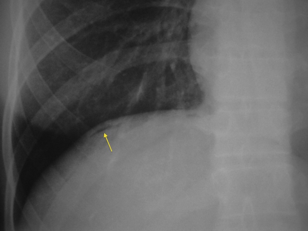
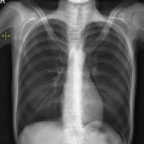
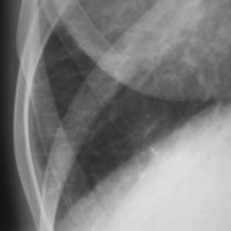
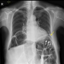
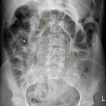
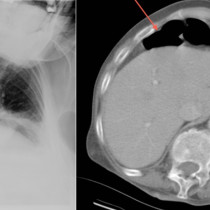
Pneumoperitoneum – erect CXR (2)

Pneumoperitoneum. This magnified view of the right hemidiaphragm from an erect CXR shows a tiny amount of free gas (arrow), showing just how sensitive this study can be.

Related Case Studies

  • Pneumoperitoneum – erect CXR
  • Pulmonary oedema
  • Perforated Duodenal Ulcer – CT
  • Chilaiditi’s syndrome with perforation
  • Pneumoperitoneum – Rigler’s sign – PFA
  • Chilaiditi Syndrome – CXR and CT
Back
  • Interventional Radiology
  • Diagnostic Imaging
    • Emergencies
      • Chilaiditi’s sign (colonic interposition)
      • Aortic dissection with haemopericardium
      • Pneumothorax on shoulder X-ray
      • Hampton’s Hump – pulmonary embolism
      • Anterior knee dislocation
      • Perilunate dislocation
      • Bilateral Perched Facets
      • Pronator quadratus fat pad sign
      • Rigler’s sign in small bowel obstruction
      • Thoracic aortic dissection – CT
      • Tracheal shift on CXR
      • Cauda Equina Compression – MRI
      • Renal infarct secondary to atrial fibrillation
      • Falciform ligament sign
      • Retroperitoneal perforation
      • Cauda equina compression
      • Small bowel obstruction – adhesions
      • Tension pneumothorax due to rib fracture
      • Chilaiditi’s syndrome with perforation
      • Lipohaemarthrosis of the knee
      • Scaphoid fracture
      • Anterior shoulder dislocation
      • Pneumothorax – expiration
      • Malpositioned NG tube
      • Large bowel obstruction
      • Femoral neck fracture
      • Salter Harris I fracture, proximal humerus
      • Triquetral fracture
      • Scaphoid fracture
      • Small bowel obstruction
      • Colitis – PFA
      • Tension pneumothorax
      • Parapneumonic effusion
      • Pleural effusions
      • Congestive cardiac failure
      • Radial head fracture
      • Radial neck fracture
      • Pulmonary oedema
      • Pneumomediastinum
      • Sigmoid volvulus
      • Small bowel obstruction
      • Pneumoperitoneum – erect CXR (2)
      • Caecal volvulus
      • Salter-Harris IV Fracture – radiograph and CT
      • Toxic megacolon – PFA/CT
      • Rigler’s sign
      • Pneumothorax post pacemaker insertion
      • Tension pneumothorax
      • Surgical emphysema
      • Pneumoperitoneum – erect CXR
      • Tension pneumothorax in CF patient
      • Pneumoperitoneum – Rigler’s sign – PFA
    • Neuro
    • Tubes & Lines
    • Trauma
    • Oncology
    • Hepatobiliary
    • Genitourinary
    • Gastrointestinal
    • Chest
    • Musculoskeletal
    • Vascular

All Case Studies

  • Main Categories

    • Interventional Radiology
      • Vascular IR
      • Non-Vascular IR
      • IR Oncology
      • Musculoskeletal IR
    • Diagnostic Imaging
      • Emergencies
      • Neuro
      • Tubes & Lines
      • Trauma
      • Oncology
      • Hepatobiliary
      • Genitourinary
      • Gastrointestinal
      • Chest
      • Musculoskeletal
      • Vascular
  • CONTACT US

    Department of Radiology
    St Vincent’s University Hospital
    Elm Park
    Dublin 4
    Send email

    Links

    • FAQs
    • Links

    SOCIAL

    © Copyright St Vincent's University Hospital Radiology Department 2025
    Web Design Dublin

    Search St. Vincent's University Hospital Radiology Department

    This website uses cookies. Accept Read More