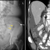
Ascites – radiograph
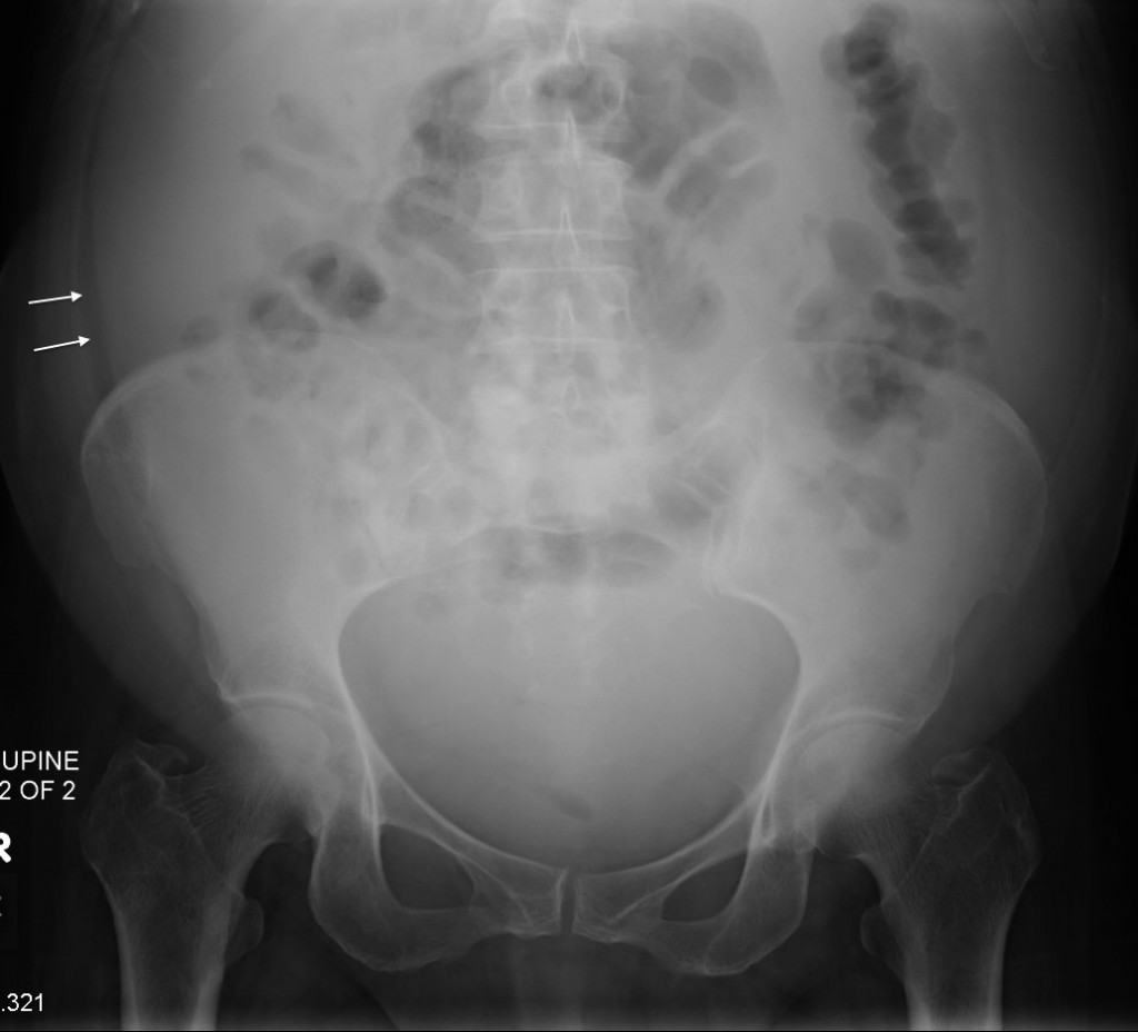
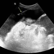
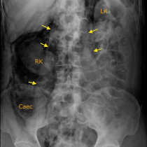
Ascites. When there is a moderate or large volume of ascites in the abdomen, we can often detect it on an abdominal radiograph. The fluid causes bulging of the flanks, as in this example, and displaces the bowel centrally, also illustrated here. A strip of fat located between the peritoneum and the abdominal wall muscles, known as the properitoneal fat stripe, becomes displaced laterally by the fluid, and is indicated by the arrows in this example. Smaller volumes of ascites are usually radiographically occult, but can easily be identified with ultrasound.