Ascites – x-ray and CT
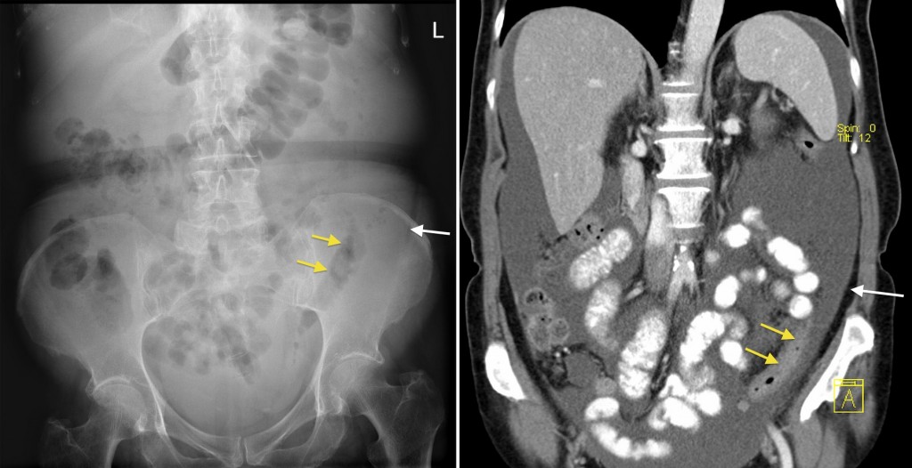
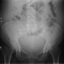
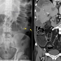
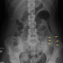
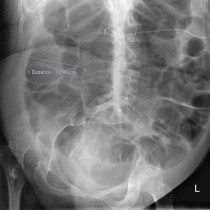
Plain radiographs are not a sensitive method of identifying ascites, as at least 500 ml of fluid is required before it can be detected. If suspected clinically, imaging confirmation is usually performed with ultrasound. Nonetheless, we will occasionally make a first diagnosis of ascites on a plain film. This case illustrates what we look for – on a supine abdominal film the fluid will tend to accumulate in the parabolic gutters. This pushes the ascending and descending colon medially, and displaces the fat deep to the abdominal wall musculature (called the properitoneal fat line) laterally. In this example, the displaced descending colon is indicated by the yellow arrows, while the displaced properitoneal fat is indicated by the white arrow. The corresponding coronal CT image gives you a better idea of what you’re seeing here – you’ll notice that there’s a large volume of fluid and that some of it is between the colon (yellow arrows) and the properitoneal fat (white arrow). There are at least 1.5-2 litres of fluid present here. The patient was subsequently diagnoses with metastatic ovarian carcinoma.