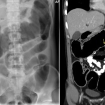
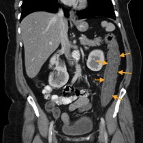
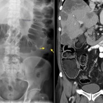
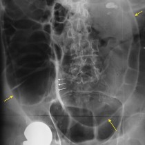
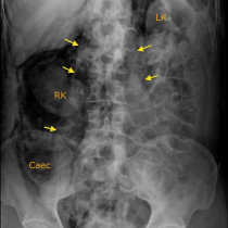
• Close
    • Home
    • Videos
    • Tutorials
    • Gallery
    • Links
    • FAQs
    • All Case Studies
    • Diagnostic Imaging
      • Imaging Modalities
        • Radiography
        • Fluoroscopy
        • Ultrasound
        • CT
        • MRI
        • Nuclear Medicine
      • Contrast Media
      • Radiological Anatomy
      • Case Studies
      • PACS
      • Radiation Safety
      • Radiological Descriptive Terms
    • Interventional Radiology
      • Introduction to IR
      • Image-guided Biopsy
      • Case Studies
    • UCD Students
      • Weekly Rotation
      • Radiology Conferences
      • Resources
      • Undergraduate Medals
    • Electives
    • Careers
    • About Us
St. Vincent's University Hospital Radiology Department
  • Diagnostic Imaging
    • Imaging Modalities
      • Radiography
      • Fluoroscopy
      • Ultrasound
      • CT
      • MRI
      • Nuclear Medicine
    • Contrast Media
    • Radiological Anatomy
    • Case Studies
    • PACS
    • Radiation Safety
    • Radiological Descriptive Terms
  • Interventional Radiology
    • Introduction to IR
    • Image-guided Biopsy
    • Case Studies
  • UCD Students
    • Weekly Rotation
    • Radiology Conferences
    • Resources
    • Undergraduate Medals
  • Electives
  • Careers
  • About Us
  • Home
  • Videos
  • Tutorials
  • Gallery
  • Links
  • FAQs
  • All Case Studies

Colitis – PFA

Colitis – PFA

Colitis. This patient with a history of ulcerative colitis presented with acute abdominal pain and bloody diarrhoea. The transverse and sigmoid colon are only mildly dilated however there is obvious severe thickening of the descending colonic wall (arrows), in keeping with colitis.

Related Case Studies

  • Toxic megacolon – PFA/CT
  • Colitis – CT
  • Large bowel obstruction
  • Sigmoid volvulus
  • Retroperitoneal perforation
  • Rigler’s sign
Back
  • Interventional Radiology
  • Diagnostic Imaging
    • Emergencies
      • Chilaiditi’s sign (colonic interposition)
      • Aortic dissection with haemopericardium
      • Pneumothorax on shoulder X-ray
      • Hampton’s Hump – pulmonary embolism
      • Anterior knee dislocation
      • Perilunate dislocation
      • Bilateral Perched Facets
      • Pronator quadratus fat pad sign
      • Rigler’s sign in small bowel obstruction
      • Thoracic aortic dissection – CT
      • Tracheal shift on CXR
      • Cauda Equina Compression – MRI
      • Renal infarct secondary to atrial fibrillation
      • Falciform ligament sign
      • Retroperitoneal perforation
      • Cauda equina compression
      • Small bowel obstruction – adhesions
      • Tension pneumothorax due to rib fracture
      • Chilaiditi’s syndrome with perforation
      • Lipohaemarthrosis of the knee
      • Scaphoid fracture
      • Anterior shoulder dislocation
      • Pneumothorax – expiration
      • Malpositioned NG tube
      • Large bowel obstruction
      • Femoral neck fracture
      • Salter Harris I fracture, proximal humerus
      • Triquetral fracture
      • Scaphoid fracture
      • Small bowel obstruction
      • Colitis – PFA
      • Tension pneumothorax
      • Parapneumonic effusion
      • Pleural effusions
      • Congestive cardiac failure
      • Radial head fracture
      • Radial neck fracture
      • Pulmonary oedema
      • Pneumomediastinum
      • Sigmoid volvulus
      • Small bowel obstruction
      • Pneumoperitoneum – erect CXR (2)
      • Caecal volvulus
      • Salter-Harris IV Fracture – radiograph and CT
      • Toxic megacolon – PFA/CT
      • Rigler’s sign
      • Pneumothorax post pacemaker insertion
      • Tension pneumothorax
      • Surgical emphysema
      • Pneumoperitoneum – erect CXR
      • Tension pneumothorax in CF patient
      • Pneumoperitoneum – Rigler’s sign – PFA
    • Neuro
    • Tubes & Lines
    • Trauma
    • Oncology
    • Hepatobiliary
    • Genitourinary
    • Gastrointestinal
    • Chest
    • Musculoskeletal
    • Vascular

All Case Studies

  • Main Categories

    • Interventional Radiology
      • Vascular IR
      • Non-Vascular IR
      • IR Oncology
      • Musculoskeletal IR
    • Diagnostic Imaging
      • Emergencies
      • Neuro
      • Tubes & Lines
      • Trauma
      • Oncology
      • Hepatobiliary
      • Genitourinary
      • Gastrointestinal
      • Chest
      • Musculoskeletal
      • Vascular
  • CONTACT US

    Department of Radiology
    St Vincent’s University Hospital
    Elm Park
    Dublin 4
    Send email

    Links

    • FAQs
    • Links

    SOCIAL

    © Copyright St Vincent's University Hospital Radiology Department 2025
    Web Design Dublin

    Search St. Vincent's University Hospital Radiology Department

    This website uses cookies. Accept Read More