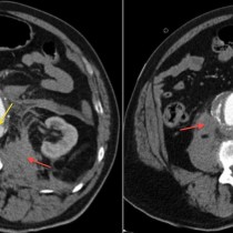
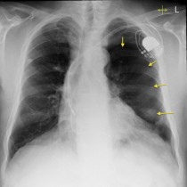
• Close
    • Home
    • Videos
    • Tutorials
    • Gallery
    • Links
    • FAQs
    • All Case Studies
    • Diagnostic Imaging
      • Imaging Modalities
        • Radiography
        • Fluoroscopy
        • Ultrasound
        • CT
        • MRI
        • Nuclear Medicine
      • Contrast Media
      • Radiological Anatomy
      • Case Studies
      • PACS
      • Radiation Safety
      • Radiological Descriptive Terms
    • Interventional Radiology
      • Introduction to IR
      • Image-guided Biopsy
      • Case Studies
    • UCD Students
      • Weekly Rotation
      • Radiology Conferences
      • Resources
      • Undergraduate Medals
    • Electives
    • Careers
    • About Us
St. Vincent's University Hospital Radiology Department
  • Diagnostic Imaging
    • Imaging Modalities
      • Radiography
      • Fluoroscopy
      • Ultrasound
      • CT
      • MRI
      • Nuclear Medicine
    • Contrast Media
    • Radiological Anatomy
    • Case Studies
    • PACS
    • Radiation Safety
    • Radiological Descriptive Terms
  • Interventional Radiology
    • Introduction to IR
    • Image-guided Biopsy
    • Case Studies
  • UCD Students
    • Weekly Rotation
    • Radiology Conferences
    • Resources
    • Undergraduate Medals
  • Electives
  • Careers
  • About Us
  • Home
  • Videos
  • Tutorials
  • Gallery
  • Links
  • FAQs
  • All Case Studies

AAA on pelvic x-ray

AAA on pelvic x-ray

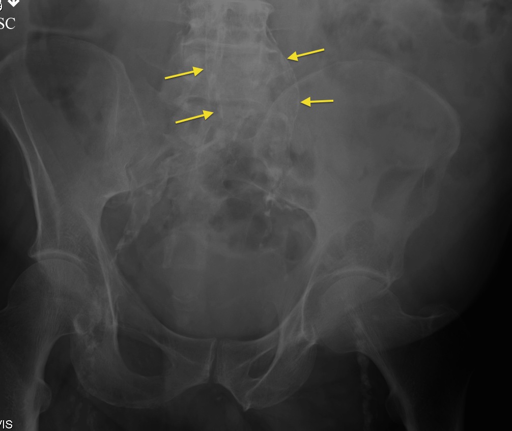
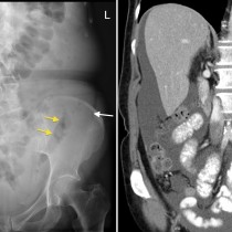
AAA. When heavily calcified, as is commonly the case in elderly patients, the walls of the aorta may be clearly visible on radiographs which can allow us to make the diagnosis of an aneurysm, as in this case (arrows). When we see a case like this, we routinely recommend an ultrasound to assess the true diameter of the aneurysm.

Related Case Studies

  • Ruptured Aortic Aneurysm
  • Deep venous thrombosis
  • Epididymo-orchitis – ultrasound
  • Pneumothorax post pacemaker insertion
  • Ascites – x-ray and CT
  • Giant Cell Arteritis – CT
Back
  • Interventional Radiology
  • Diagnostic Imaging
    • Emergencies
    • Neuro
    • Tubes & Lines
    • Trauma
    • Oncology
    • Hepatobiliary
    • Genitourinary
    • Gastrointestinal
    • Chest
    • Musculoskeletal
    • Vascular
      • Ruptured aneurysm mimicking renal colic
      • Calcified abdominal aortic aneurysm – lumbar spine radiograph
      • Thoracic aortic dissection – CT
      • Vasculitis – PET
      • Giant Cell Arteritis – CT
      • Carotid Stenosis
      • Arterial calcification in a diabetic
      • AAA on pelvic x-ray
      • Deep venous thrombosis
      • Ruptured Aortic Aneurysm

All Case Studies

  • Main Categories

    • Interventional Radiology
      • Vascular IR
      • Non-Vascular IR
      • IR Oncology
      • Musculoskeletal IR
    • Diagnostic Imaging
      • Emergencies
      • Neuro
      • Tubes & Lines
      • Trauma
      • Oncology
      • Hepatobiliary
      • Genitourinary
      • Gastrointestinal
      • Chest
      • Musculoskeletal
      • Vascular
  • CONTACT US

    Department of Radiology
    St Vincent’s University Hospital
    Elm Park
    Dublin 4
    Send email

    Links

    • FAQs
    • Links

    SOCIAL

    © Copyright St Vincent's University Hospital Radiology Department 2025
    Web Design Dublin

    Search St. Vincent's University Hospital Radiology Department

    This website uses cookies. Accept Read More