Spinal cord metastasis – MRI
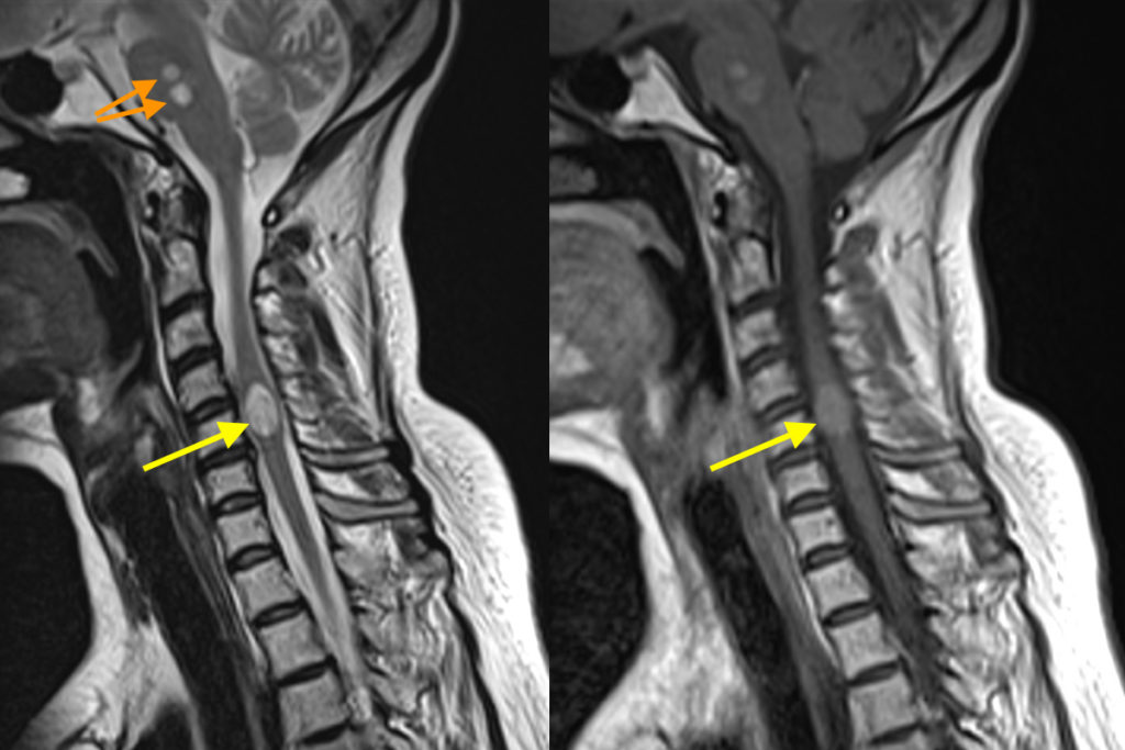
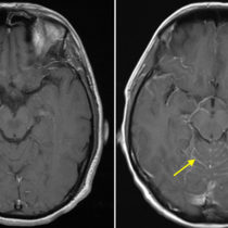
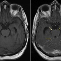
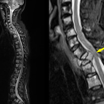
These MR images are from a patient with metastatic lung cancer and known brain metastases who presented with progressive lower limb weakness. The image on the left is from the sagittal T2-weighted sequence (note the bright CSF) and shows a hyperintense lesion in the cervical spinal cord (yellow arrow). Note how the lesion is also causing expansion of the cord. On the right is the corresponding sagittal T1-weighted post-gadolinium image where you can see that the lesion enhances. There are additional metastases in the brainstem (orange arrows).
Although there is a differential diagnosis for this imaging appearance (for example, primary spinal cord neoplasms and demyelination), in a patient with this history we can confidently call this a metastasis.
In most patients with metastatic disease in the spine, the metastases involve the vertebrae. They may end up causing cord compression when they extend through the vertebral body wall into the spinal canal, or can cause cord compression if the vertebra fractures as a result of the metastasis. The next most common location to encounter spinal metastases is the leptomeninges. This results from seeding through the CSF. Spinal cord metastases are relatively rare, and are usually encountered in patients who already have extensive metastatic disease elsewhere. They will often have additional metastases in the brain, as in this example. The most common primary malignancy to metastasize to the spinal cord is lung cancer, however it can also occur in patients with breast cancer, malignant melanoma, lymphoma and renal cell cancer.
Patients with cord metastases typically present with weakness and/or paraesthesia. When suspected, a contrast-enhanced MRI is necessary – cord metastases are almost never visible on CT. MRI will clearly demonstrate all forms of spinal metastases – vertebral, leptomeningeal and cord lesions. The vertebral lesions will be visible on non-contrast MRI however most leptomeningeal metastatic disease and some cord metastases will only be apparent on a post-contrast MRI. See an example of leptomeningeal metastatic disease here.