Polyostotic Paget’s disease
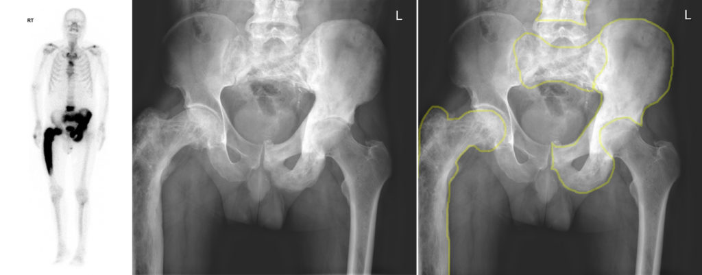
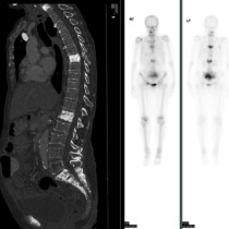
This 65-year-old man presented with thigh pain, and was referred for hip radiographs on the basis of which a whole-body bone scan was performed.
The bone scan, left, shows greatly increased uptake in the right femur, which appears bowed when you compare it to the left, as well in as the left hemipelvis, sacrum and several thoracic and lumbar vertebrae.
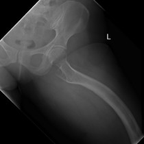
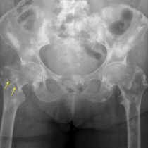
The pelvic radiograph shows increased density of the affected bones, outlined in yellow on the right-hand image. You will also notice that the cortex of the right femur is thicker than on the left, and the trabecular markings within it are much more prominent than on the opposite side. The femur also appears expanded.
These radiographic appearances, and the corresponding increased uptake on the bone scan, are characteristic of Paget’s disease. This is a fairly common condition that usually affects patients over the age of 55, and is more common in men. The disease causes increased bone turnover in the affected parts of the skeleton, resulting in softer bones which can become deformed (like the right femur in this case) and which are also prone to fractures. However, in many patients Paget’s disease is asymptomatic and is picked up as an incidental finding on imaging.
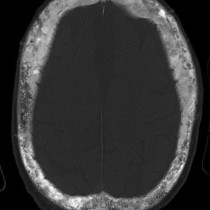
In some patients, only one bone is involved, while in others it can involves multiple sites. In the latter case, this is called ‘polyostotic Paget’s disease’. The most commonly involved bones are the femur, ilium, sacrum, vertebrae and skull, although any bone can be affected.
When long bones like the femur are involved and become bowed, this results in abnormal stresses through adjacent joints therefore patients are prone to development of osteoarthritis. A relatively rare complication of Paget’s disease is malignant transformation, most frequently into osteosarcoma. Involvement of the skull can result in stenosis of the skull base foramina and compression of the cranial nerves passing through them.