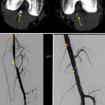
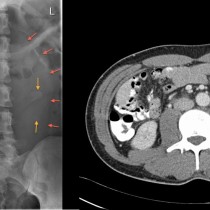
Pelvic embolization
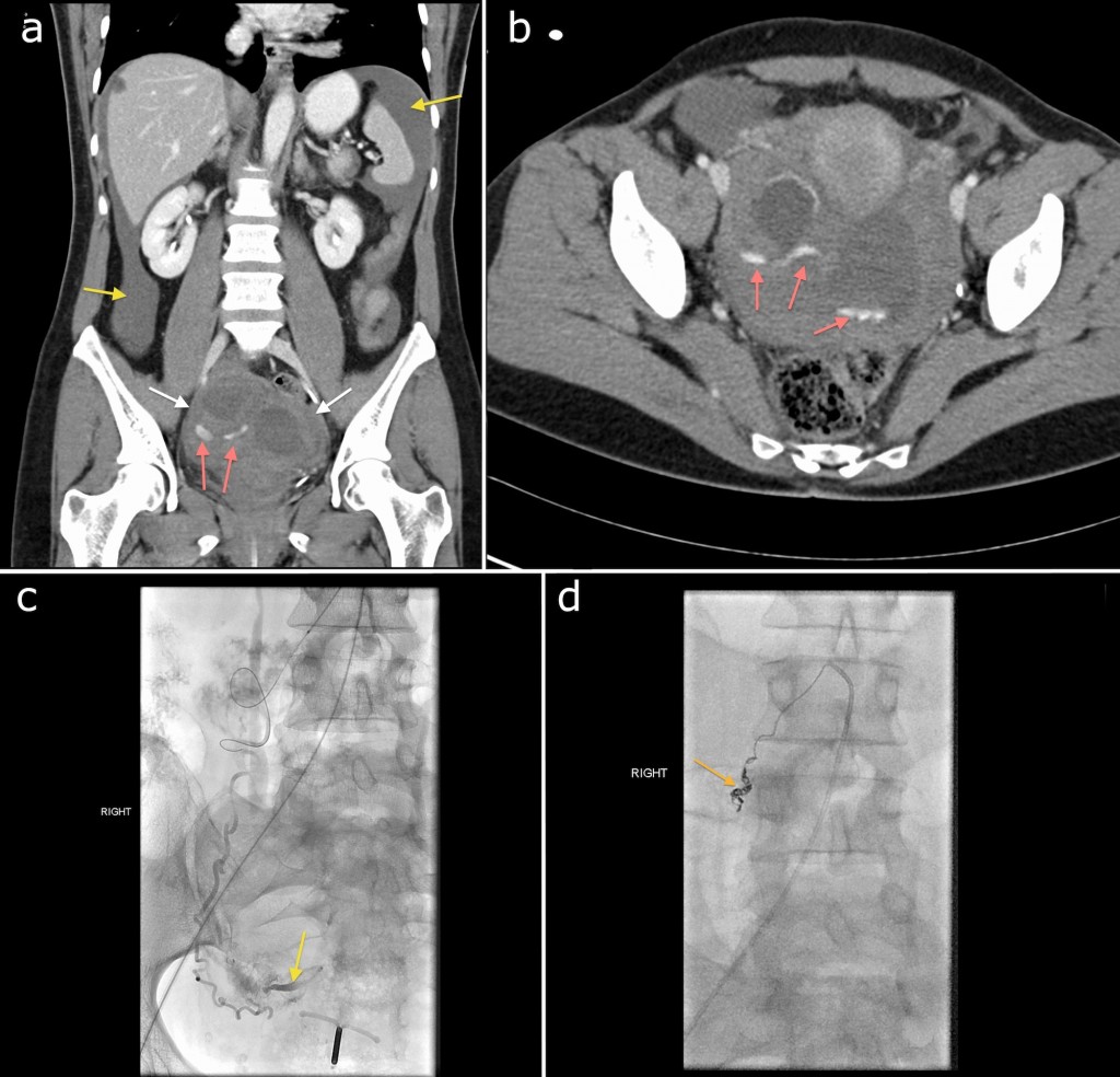
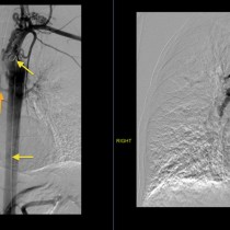
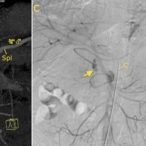
This is an example of a life-saving Interventional Radiology procedure. This young woman presented with acute abdominal pain and quickly became haemodynamically unstable. CT showed haemoperitoneum (yellow arrows) with evidence of extravasation of IV contrast into the right side of the pelvis (the pink arrows on both CT images show linear areas of contrast extravasation), indicating active haemorrhage. She was brought straight to IR, by which stage her haemoglobin was 5 and her systolic pressure 60. Angiography, (c), showed that the source of the haemorrhage was the right ovarian artery (the yellow arrow on this image shows contrast extravasation at the same site as was demonstrated on the CT). The ovarian artery was embolized with coils (metallic density on image (d), arrow). The patient stabilised and the haemorrhage did not recur. No underlying pelvic abnormality was identified and it was assumed that the source of the haemorrhagic in this case was a ruptured ovarian cyst – these can occasionally be associated with severe haemorrhage.