Pancreatic pseudocyst
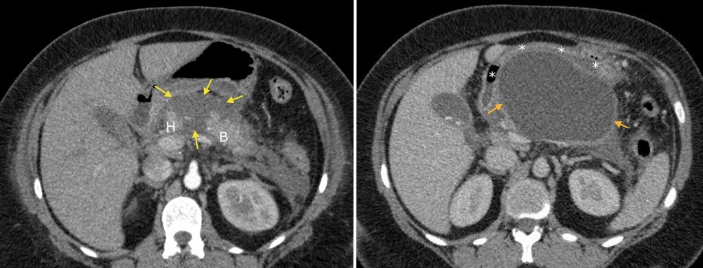
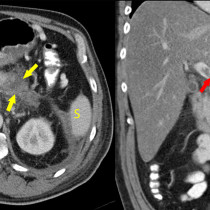
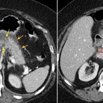
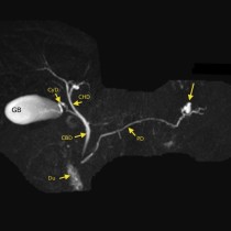
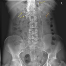
This young woman presented with acute gallstone pancreatitis. Initial CT, left, shows normal density of the pancreatic head (H) and lateral aspect of the body (B), however the large area of low density between them (yellow arrows) represents pancreatic necrosis. She represented 6 weeks later with nausea and upper abdominal pain and follow-up CT showed a 12 cm thick-walled pseudocyst extending from the site of previous necrosis into the lesser sac (orange arrows). Note the mass effect on the stomach, which is stretched around the anterior aspect of the pseudocyst (indicated by the asterisks). Also note the calcified gallstones in the gallbladder.
This is the typical location for pancreatic pseudocysts to develop, however they can be found anywhere around the pancreas or, commonly enough, quite remote from the pancreas – for example, in the pelvis or in the mediastinum.