Acute pancreatitis
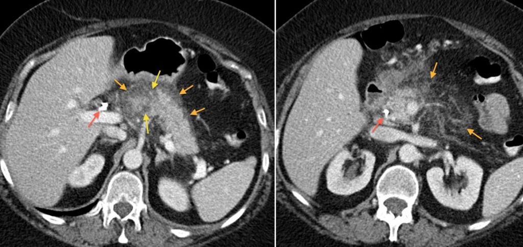
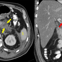
Acute pancreatitis. CT is the standard imaging modality when assessing patients with suspected acute pancreatitis, both for confirmation of the diagnosis and for follow-up during treatment. This patient developed pancreatitis due to common bile duct calculi impacted at the ampulla, for which she required ERCP and a CBD stent (indicated by the red arrows in these images). The image on the left shows inflammatory stranding of the fat around the pancreas, in addition to some peripancreatic fluid (orange arrows). While the majority of the pancreas enhances normally, there are some areas that are abnormally low in attenuation (indicated by the yellow arrows), which represent pancreatic necrosis. The right-hand image shows further ‘fat stranding’ extending from the pancreas into the small bowel mesentery (orange arrows).