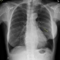
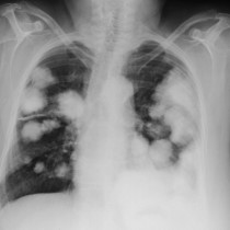
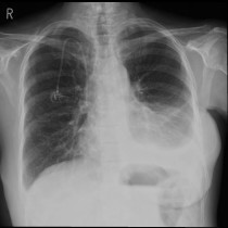
Mastectomy – CXR
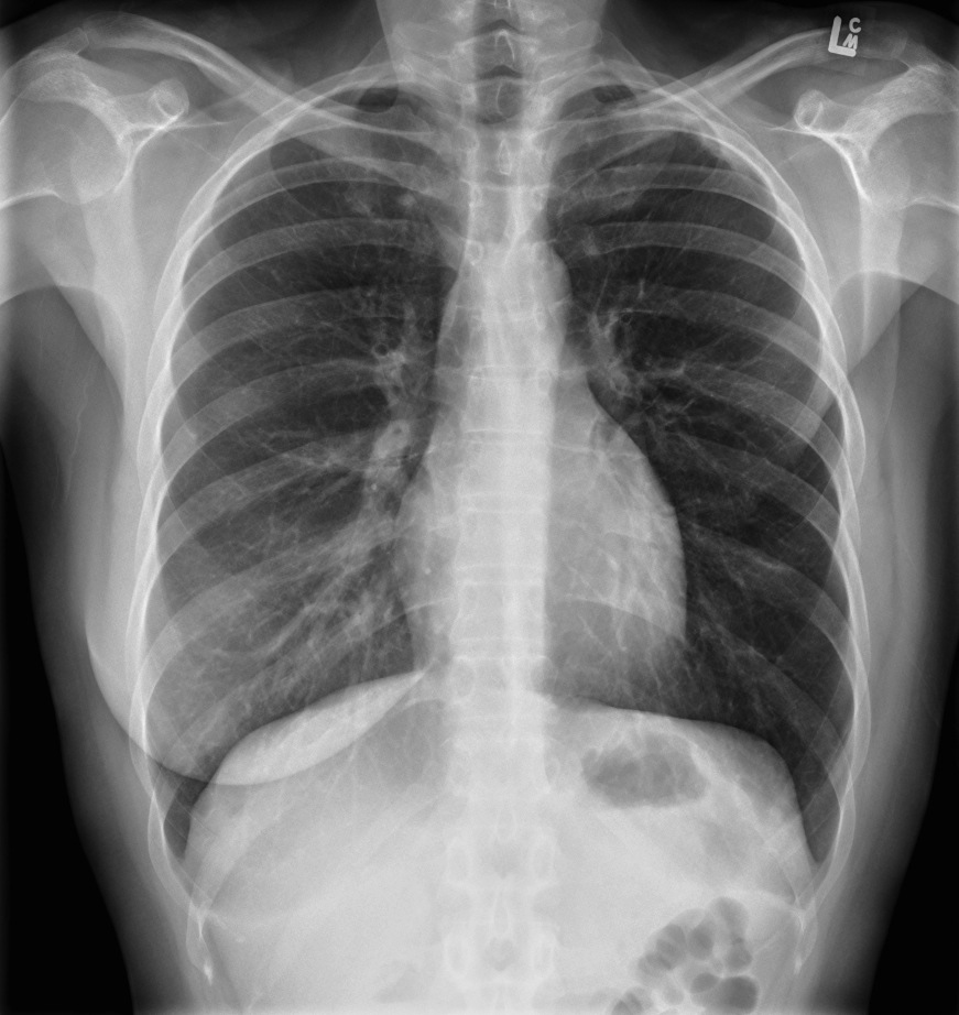
Mastectomy. This patient has had a previous left mastectomy. Note how the left lung appears relatively hyperlucent when compared to the right, because of the reduced soft tissue between the x-ray tube and detector. It’s fairly obvious in this example, but occasionally it can be difficult to spot that there has been a previous mastectomy, which can trick us into thinking that the asymmetry of the lungs is due to a pathological process. It’s vital that you check the breast shadows for symmetry in every female patient’s CXR. Remember, in an exam situation, when you see evidence of a mastectomy (or you see clips in the breast from a wide local excision) keep looking around for evidence of metastatic disease – in breast cancer, this can involve the lungs, pleura (producing an effusion), hilar/mediastinal nodes and bones (breast cancer bones mets are usually sclerotic).