Mandibular fractures – OPG
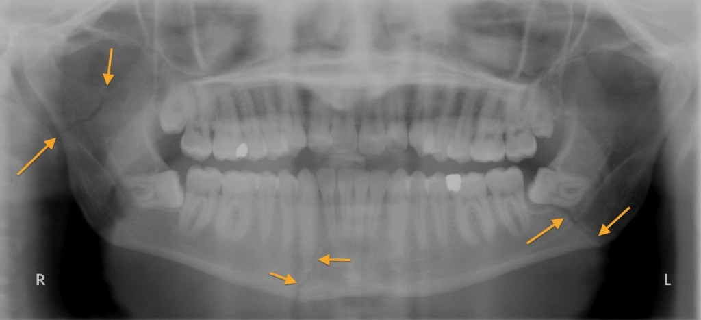
This image is called an OPG – short for orthopantomogram. This technique uses a special type of x-ray equipment that allows a rotational, panoramic image of the facial bones and jaw to be obtained, which gives us a beautiful view of the entire mandible and both temporomandibular joints (TMJs). In SVUH we typically use this in the setting of trauma looking for fractures and dislocations, although it is sometimes also requested by the dental service when they want to assess for dental caries.
This young man was punched in the jaw and presented to ED several days later with persistent pain and difficulty chewing. The image shows three fractures, indicated by the arrows. The mandible has a tendency to fracture in more than one place, which is something it has in common with several other bony structures around the body, This is due to what is known as the ‘ring-bone rule’. Although the mandible isn’t exactly ring-shaped, it acts like any ring-shaped structure because it is relatively fixed at the TMJs. Structures that are subject to the ring-bone rule tend to break in at least two places. An analogy is to imagine a polo mint – try breaking one of these in one place only, and you will find it impossible. Other places where this rule applies include the atlas (C1 vertebra), which is truly ring-shaped, and the pelvic bones (for example, fractures of the superior pubic ramus are almost always accompanied by fractures of the inferior ramus).