Kienbock’s disease
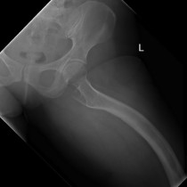
This woman in her 40s presented with progressive wrist pain.
She had a history of a previous severe wrist injury, years previously, where she suffered a lunate dislocation (see an example of this here), for which she underwent surgical reduction.
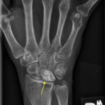
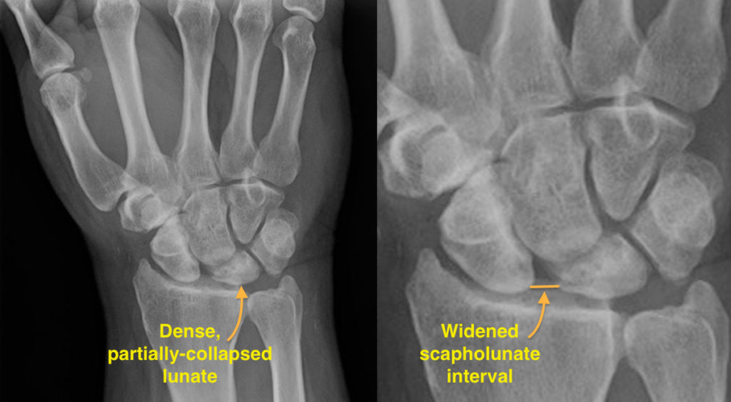
This radiograph shows increased density of the lunate, which also has an abnormal shape due to partial collapse. These are relatively advanced features of avascular necrosis (AVN), also known as Kienbock’s disease when it occurs in the lunate.
Kienbock’s disease can occur spontaneously, but is also a recognized complication of previous wrist trauma as in this case. It is also associated with ‘negative ulnar variance’, where the ulna is slightly shorter than the radius (by greater than 2 mm). The exact mechanism of this association is not yet known but it is presumed to be due to altered blood supply to the lunate.
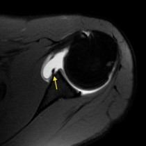
MRI is more sensitive in the earlier stages of lunate AVN, showing abnormal marrow signal before any radiographic changes occur.
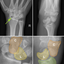
In this example, there is widening of the distance between the scaphoid and lunate, which indicates that the scapholunate ligament is torn – this was a consequence of the previous dislocation and, even in isolation, would predispose to early onset of osteoarthritis at the wrist.
If you are like I was as a medical student (and on Monday mornings) and are rusty with your carpal bone anatomy, you can revise this here.