Perilunate dislocation
This young man presented to the ED after a fall on his outstretched hand (usually termed a ‘FOOSH’ by the ED docs).
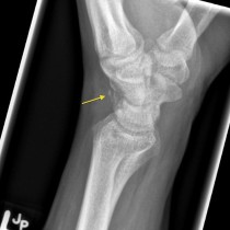
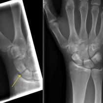
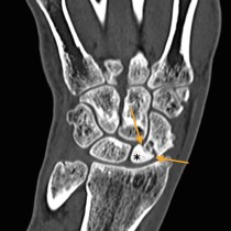
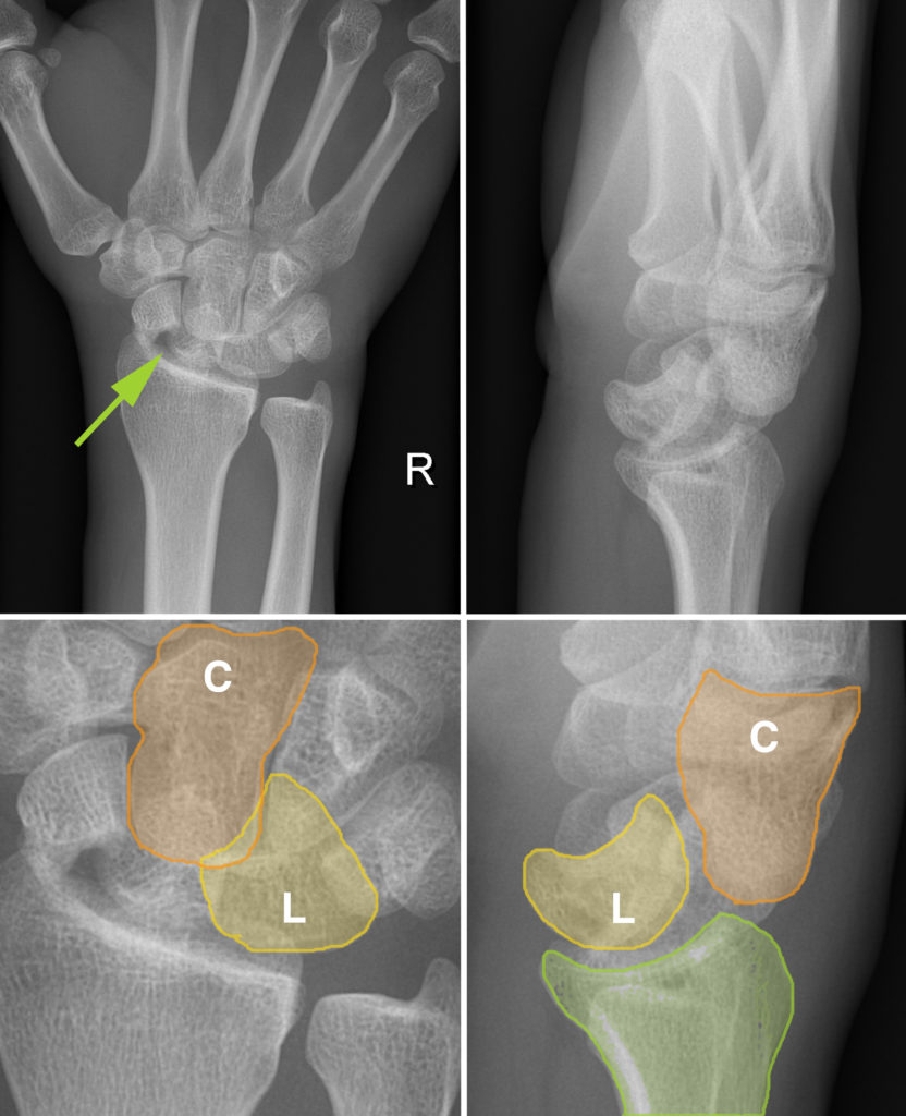
At first glance, the AP radiograph of his right wrist (top left image) appears to show a solitary abnormality – a displaced fracture of the scaphoid waist (arrow). However, a much more serious injury is present. If you look carefully, you will notice that there is slightly too much overlap of the proximal carpal row (scaphoid, lunate and triquetral) with one of the bones in the distal carpal row (the capitate). This is better appreciated on the magnified view (below left), with the capitate (C) highlighted in orange, and the lunate (L) in yellow. Also, the shape of the lunate is unusual for an AP radiograph – it looks a little bit like a slice of pie (you can review the normal appearance of the carpal bones here).
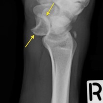
These findings should make us suspicious of a carpal dislocation, which the lateral radiograph (top right) confirms. This shows abnormal alignment of the capitate relative to the lunate. Again, the magnified view below makes this easier to understand. The capitate is dislocated dorsal to the lunate, and its position explains the abnormal overlap that we see on the AP. The dislocated capitate pushes and tilts the lunate forward slightly which is why it ends up looking ‘pie-shaped’ on the AP view.
This injury looks very similar to another type of carpal dislocation – lunate dislocation. The key difference between these two entities is their appearance on the lateral radiograph. In perilunate dislocation, as in this case, the lunate remains sitting in the concave articular surface of the distal radius (highlighted in green here). In lunate dislocation, the capitate and distal radius are aligned normally, but the lunate is shifted forward – see an example of this here.
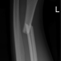
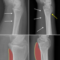
Another big difference between perilunate and lunate dislocation is that perilunate dislocation is associated with a fracture in around 75% of cases. While this fracture can involve any of the carpal bones, in the majority of cases it affects the scaphoid, like in this example. This combination of injuries is known as ‘trans-scaphoid perilunate dislocation’.
When no fracture is present, perilunate and lunate dislocations can be very subtle radiographically – unfortunately, these injuries are frequently missed initially and the diagnosis may be significantly delayed as a result. They generally necessitate open reduction with ligament reconstruction.