Pronator quadratus fat pad sign
This can be a very useful sign when evaluating wrist radiographs for a distal radial fracture. These fractures are usually easy enough to spot but can occasionally be quite subtle, especially in young adults where they are often minimally displaced.
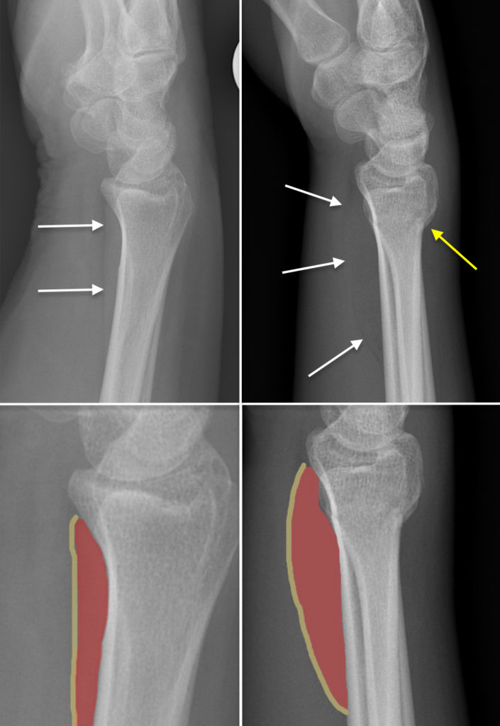
In the normal wrist, the lateral view will show a stripe of soft tissue density anterior to the distal radius which is caused by the pronator quadratus muscle. Immediately anterior to this, separating it from the flexor muscles, there is a thin lucent stripe of fat called the pronator fat pad.
When the distal radius is injured, haemorrhage around the fracture can displace the pronator muscle anteriorly which results in displacement and bulging of the fat pad. Although not very reliable, this sign has been reported to be present in up to 80% of distal radial fractures1. When we’re unsure whether to call a dubious abnormality in the radius a fracture, finding a displaced pronator fat pad may be enough to make a more confident diagnosis of fracture.
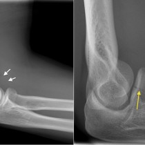
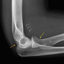
The example on the top left here is from a normal lateral wrist radiograph. The fat pad is indicated by the white arrows. On the zoomed image on the bottom left, the pronator quadratus muscle is coloured red and the fat pad in yellow.
The top right image is from a patient in his twenties who fell on his outstretched hand. There is a subtle fracture, with some buckling of the posterior cortex of the distal radius indicated by the yellow arrow. However, any doubt about the fracture is removed by the presence of a markedly displaced and bowed pronator fat pad. Again, a zoomed version is shown on the bottom right with the muscle and fat pad shadowed in red and yellow.
Reference:
- Fallahi F. Explorative study of the sensitivity and specificity of the pronator quadratus fat pad sign. Skeletal Radiology 2013. doi: 10.1007/s00256-012-1451-0