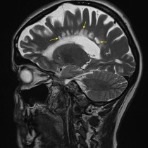
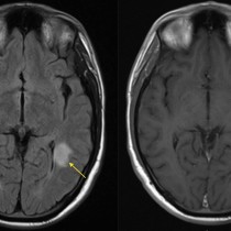
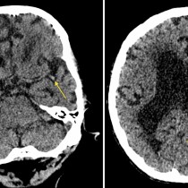
Dural AVM
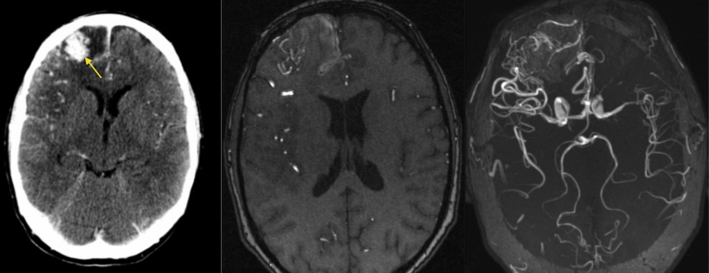
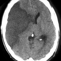
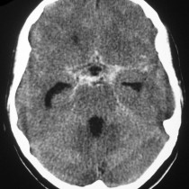
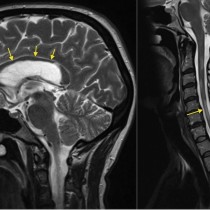
Arteriovenous malformation. This young man presented with acute onset headache. His contrast-enhanced CT, left, shows a large enhancing mass in the right frontal region (arrow). You will also notice that there are multiple prominent blood vessels in this region. MR angiography was performed to further evaluate this dural AVM; the middle image shows the abnormal vessels feeding and draining the AVM, while the right-hand image is a ‘MIP’ (maximum intensity projection) which combines multiple slices (a 3D volume of the patient) into a single 2D image. You can read more about MIPs, which are commonly used in CT, MR and PET imaging, here. Cerebral AVMs can be picked up as incidental findings, or may present with seizures, headaches, acute haemorrhage or ischaemia.