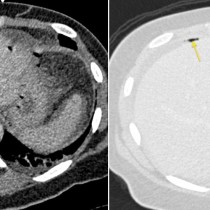
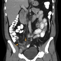
Drug paraphernalia in vagina
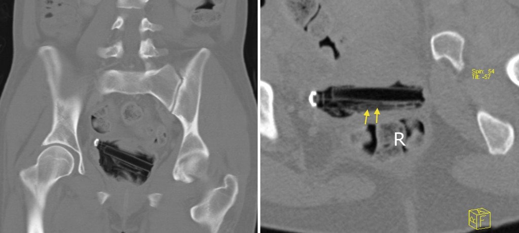
This is not something you’ll see every day. This woman, an IV drug user, was admitted through the ED with a groin abscess. CT performed to assess the extent of the abscess showed that she had come prepared – her vagina contains a dismantled syringe as well as a small bag of heroin (right-hand image, arrows). R = rectum.