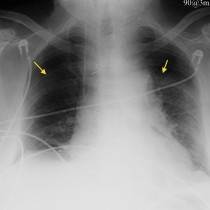
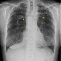
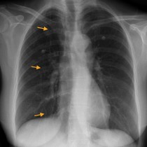
Defibrillation pads – CXR
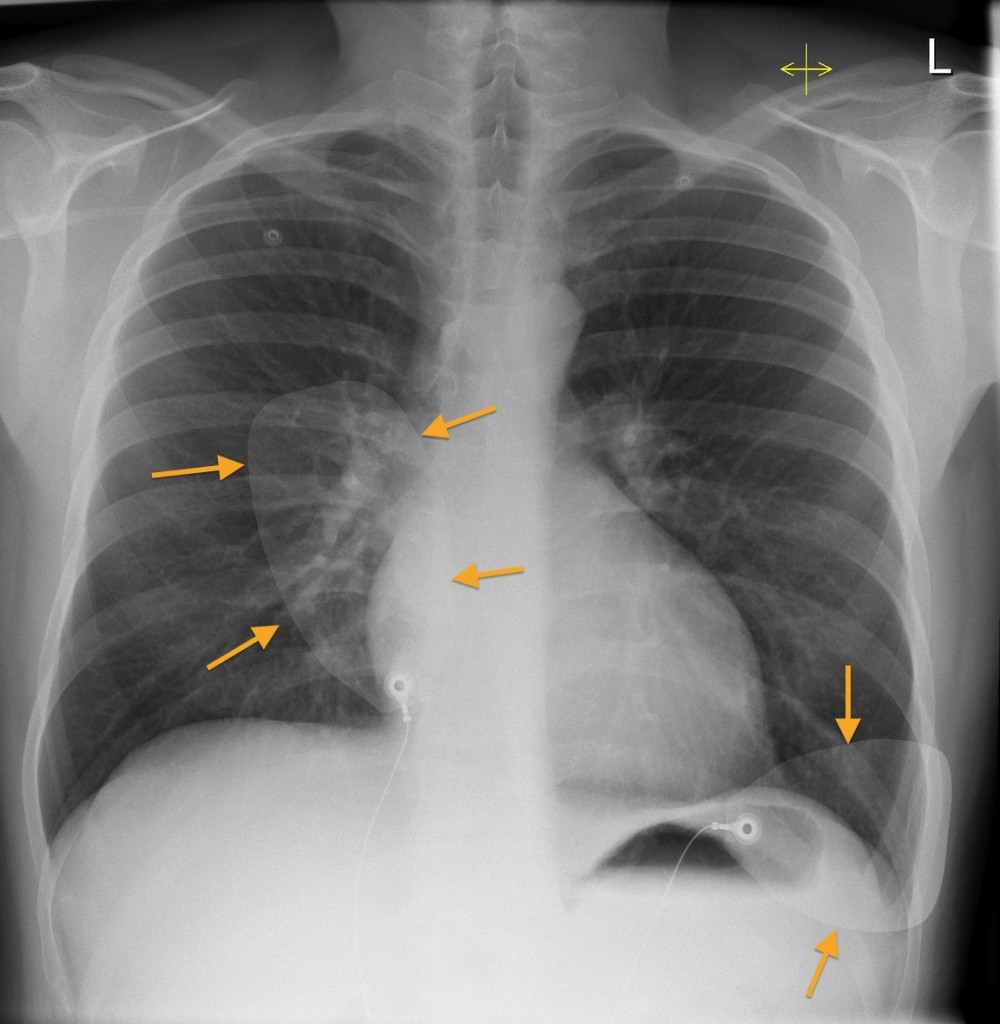
Defibrillation pads. These pads, indicated by the arrows, obviously indicate that we are dealing with a very sick patient. In addition to a careful search for signs of pulmonary oedema, bear in mind that the patient may well have required CPR prior to the radiograph being taken, so check for iatrogenic rib fractures and pneumothoraces.