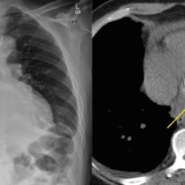
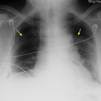
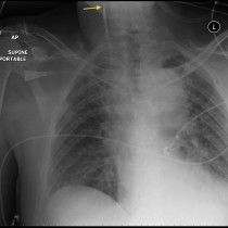
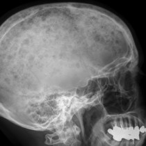
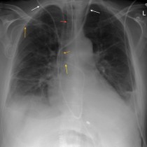
Ventriculoperitoneal shunt
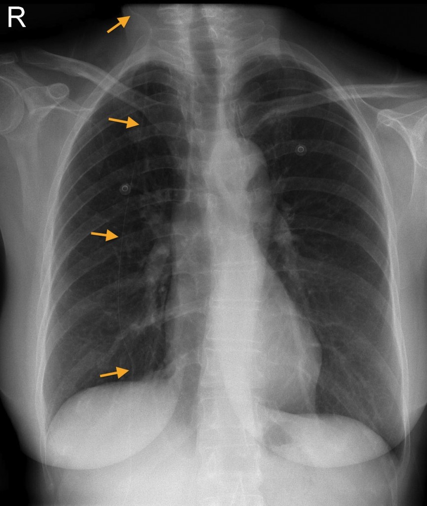
VP shunt. If you look carefully, you can a thin catheter extending from the top of this patient’s CXR through the neck and chest to the upper abdomen (arrows). This is a ventriculoperitoneal shunt, used to treat hydrocephalus. When shunt dysfunction is suspected, we are often asked to perform a radiographic ‘shunt series’, which comprises a lateral skull radiograph, frontal CXR and PFA to demonstrate the full length of the shunt and identify any discontinuity in the catheter.