Calcaneal Fracture
The calcaneus is the most commonly fractured tarsal bone, and unfortunately calcaneal fractures have a relatively poor prognosis as they often involve one or more of its articular surfaces and may be associated with additional soft tissue injuries, such as damage to the adjacent peroneal tendons.
The typical history is of a fall from a height, landing on the feet – as a result, bilateral calcaneal fractures are quite common. Concomitant lumbar spine fractures are present in up to 10% of patients with calcaneal fractures.
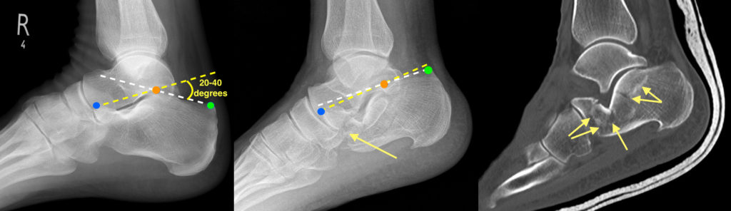
These fractures are usually visible on radiographs, however an additional radiographic tool that is helpful when trying to assess the calcaneus is to measure Bohler’s angle. This is the angle formed between two lines, one drawn between the most superior point of the anterior process of the calcaneus (the blue dot on these images) and the most superior aspect of the posterior articular surface of the calcaneus (orange dot), and the other line drawn between the latter and the most superior aspect of the calcaneal tuberosity (green dot). The angle between the two lines should normally be between 20 and 40 degrees. If it less than 20 degrees, it implies that the calcaneus has been flattened – this finding has 99% specificity for a fracture.
In this example, the image on the left is from a normal foot and shows a normal Bohler’s angle. In the middle image, from a patient who fell off a high wall and landed on his feet, the radiograph shows Bohler’s angle to be close to 0 degrees. You can see a fracture in the anterior part of the calcaneus (arrow). However, calcaneal fractures are routinely assessed with CT as radiographs often underestimate the extent of the fracture – what appears to be a simple extra-articular calcaneal fracture on a radiograph will often turn out to be severely comminuted with involvement of the joint surfaces on CT, therefore it is a vital tool for orthopaedic surgeons planning surgery for these patients. In this case, multiple fracture lines are visible on the CT image, right (arrows).