Bilateral Perched Facets
This is an uncommon cervical spine injury that results from hyperflexion of the neck.
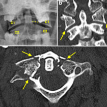
The normal facet joints in the spine are formed by the articulation of the inferior articular process of the upper vertebra and the superior articular process of the vertebra below it. In facet dislocation, the inferior articular processes of one vertebra become displaced anteriorly relative to the superior articular processes below. If there is still some contact between the articular processes, as in this example, the injury is called a ‘perched’ facet. With further anterior movement, the inferior articular process can end up anterior to the superior articular process, i.e. completely dislocated, which is called a ‘locked’ facet. These injuries can be unilateral or bilateral. As you would expect, anterior dislocation of the facets results in abnormal alignment of the relevant vertebral bodies too.
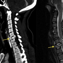
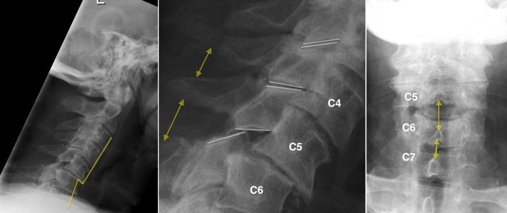
In this case, the C5-C6 facets are perched bilaterally. As a result, there is disruption of the anterior spinal line (line on the left-hand image). If you are unfamiliar with the concept of assessing spinal lines on a lateral cervical spine x-ray, have a look here.
The abnormal facet alignment is illustrated by the white lines on the middle image, which shows the normal appearance of the facet joints at the two levels above the injury.
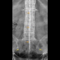
These injuries are commonly associated with other radiographic abnormalities. Tearing of the posterior ligament complex can result in widening of the space between the spinous processes at the level of the dislocated facet joints (yellow arrows – compare with the distance between the spinous processes at the level above). This is also visible on the AP view (arrows).
When facet joint injuries are unilateral, there is usually some rotation of the affected vertebra. This can be detected on the AP view, where the affected spinous process is displaced to one side (the side of the injury).
If the joint is fully dislocated, on the lateral view the affected vertebra will be displaced anteriorly by around 25% of its AP diameter. This causes some stenosis of the spinal canal but in most patients the spinal cord is unaffected.
Conversely, in bilateral facet dislocation, the vertebral body shifts forward by around 50% of its diameter, and spinal cord injury is much more common. In the case presented here, which is at the more severe end of the spectrum of perched facet joints, the C5 vertebral body has subluxed anteriorly by around 25% of its AP diameter. The patient had no neurologic deficit.