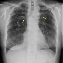
Tension pneumothorax in CF patient
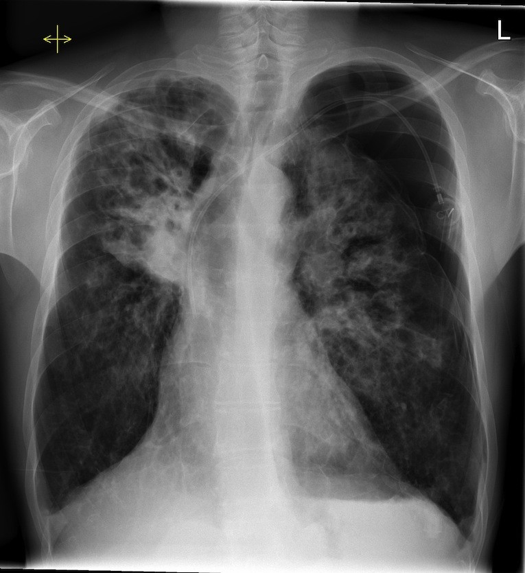
Tension pneumothorax. This patient has severe changes of cystic fibrosis – note the fibrotic change and bronchiectasis in the mid and upper zones of both lungs. One of the potential acute complications of CF is a pneumothorax, which should be carefully searched for in any CF patient who suddenly deteriorates. This is a left-sided tension pneumothorax – the trachea and the heart are shifted to the right, and there is increased lucency in the left upper zone with absent lung markings.
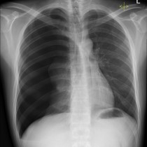
The extent of the chronic changes in the lungs in patients with severe CF often make it difficult to determine whether or not there is any superimposed consolidation. Comparison with a previous CXR (of which there are usually plenty) is vital for this reason, to spot subtle differences between the two.
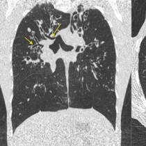
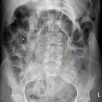
As you will be aware, routine imaging of CF patients includes high resolution CT chest to assess for progression of bronchiectasis and fibrosis, as well as abdominal ultrasound to monitor the potential intra-abdominal manifestations of the disease (such as cirrhosis, portal hypertension, pancreatic cystosis).
Note also the portacath on the left in this example. Whenever you see one of these on a CXR, you are dealing with a patient who is in need of long-term intravenous therapy and should think about underlying malignancy as well as the possibility of an underlying condition that requires long-term or repeated courses of antibiotics, particularly cystic fibrosis.