Ruptured pericallosal aneurysm with subarachnoid haemorrhage – CT
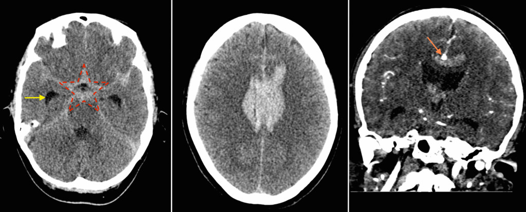
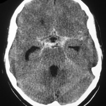
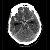
The non-contrast CT brain image on the left shows the typical appearance of a large acute subarachnoid haemorrhage – the high-attenuation 5-pointed star (outlined in red) centred on the circle of Willis. In this case, the temporal horns of the lateral ventricles are already dilated (yellow arrow), indicating that hydrocephalus has developed, which is one of the potential complications of subarachnoid haemorrhage.
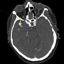
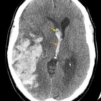
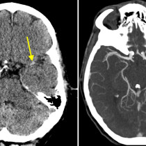
In this example, there was a very large quantity of acute blood located several slices above the circle of Willis, draped over the corpus callosum (middle image). This finding suggested that the ruptured aneurysm (assuming that an aneurysm was the cause of the subarachnoid haemorrhage) was located in this region. Subsequent CT angiogram confirmed that this was indeed the case. The coronal CT angiogram image on the right shows the round aneurysm (orange arrow) in the midline, immediately above the corpus callosum. This aneurysm was arising from the right pericallosal artery, which is the continuation of the anterior cerebral artery.
As a reminder, the causes of subarachnoid haemorrhage include:
- Trauma
- Ruptured berry aneurysm (which account for 85% of non-traumatic subarachnoid haemorrhage and occur with higher incidence in patients with adult polycystic kidney disease, Ehlers-Danlos syndrome, Osler-Weber-Rendu syndrome, neurofibromatosis and other conditions)
- Cerebral or spinal arteriovenous malformation
- Vasculitis
- Coagulopathy
- Cerebral venous thrombosis/venous infarction
For a review of the CT appearances of various types of intracranial haemorrhage, go to the tutorials section where you find a slideshow presentation on acute intracranial pathology.