Radiofrequency ablation – HCC
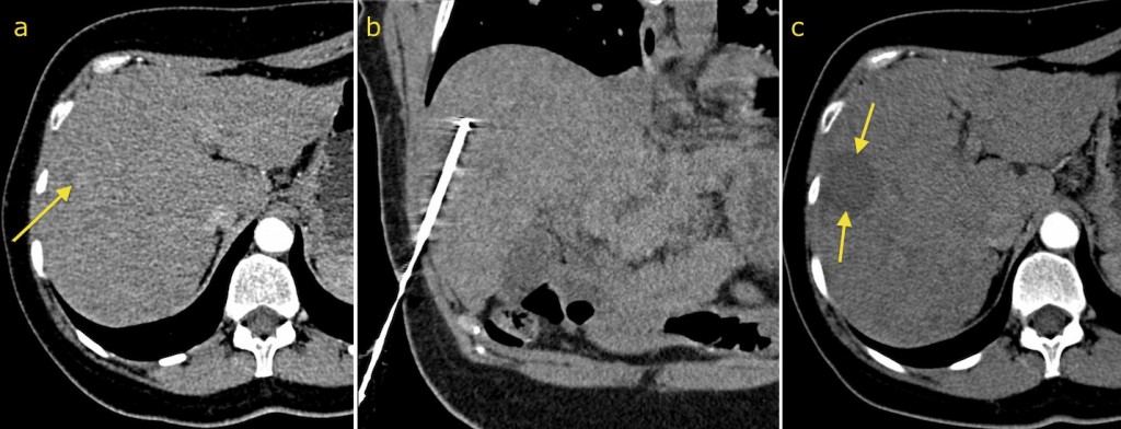
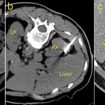
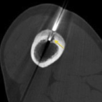
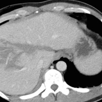
Radiofrequency ablation – hepatocellular carcinoma. The rather subtle small focus of hypervascularity, arrow on image (a), in the liver of this 51 year old woman with chronic liver disease showed typical CT characteristics of hepatocellular carcinoma. Radiofrequency ablation was requested. This is performed under CT guidance, and is one of the few IR procedures that requires general anaesthesia. The radiofrequency probe is advanced through the liver so that its tip is in the centre of the tumour, image (b). Ablation is performed by connecting the probe to a radiofrequency device which heats its tip to a high temperature which causes the death of cells in a sphere around it. The size of this ablation sphere can be varied by using probes with different sizes of active tips. Follow-up CT (c) shows no evidence of residual hypervascular HCC; instead, at the site of the previous tumour there is a sphere of low density, arrows, representing the ‘ablation zone’.