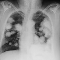
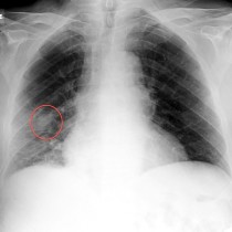
• Close
    • Home
    • Videos
    • Tutorials
    • Gallery
    • Links
    • FAQs
    • All Case Studies
    • Diagnostic Imaging
      • Imaging Modalities
        • Radiography
        • Fluoroscopy
        • Ultrasound
        • CT
        • MRI
        • Nuclear Medicine
      • Contrast Media
      • Radiological Anatomy
      • Case Studies
      • PACS
      • Radiation Safety
      • Radiological Descriptive Terms
    • Interventional Radiology
      • Introduction to IR
      • Image-guided Biopsy
      • Case Studies
    • UCD Students
      • Weekly Rotation
      • Radiology Conferences
      • Resources
      • Undergraduate Medals
    • Electives
    • Careers
    • About Us
St. Vincent's University Hospital Radiology Department
  • Diagnostic Imaging
    • Imaging Modalities
      • Radiography
      • Fluoroscopy
      • Ultrasound
      • CT
      • MRI
      • Nuclear Medicine
    • Contrast Media
    • Radiological Anatomy
    • Case Studies
    • PACS
    • Radiation Safety
    • Radiological Descriptive Terms
  • Interventional Radiology
    • Introduction to IR
    • Image-guided Biopsy
    • Case Studies
  • UCD Students
    • Weekly Rotation
    • Radiology Conferences
    • Resources
    • Undergraduate Medals
  • Electives
  • Careers
  • About Us
  • Home
  • Videos
  • Tutorials
  • Gallery
  • Links
  • FAQs
  • All Case Studies

Primary lung cancer

Primary lung cancer

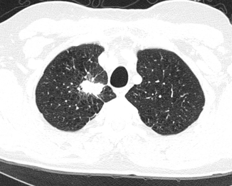
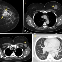
Lung cancer. The typical appearance of a primary lung cancer on CT is a soft tissue mass with irregular (‘spiculated’) margins, as illustrated in this example. In contrast, lung metastases are usually smooth and rounded in appearance on CT.

Related Case Studies

  • Brain metastases
  • Metastatic breast cancer
  • Lung metastases
  • Sclerotic Bone Metastases
  • Lung metastases (2)
  • Lung cancer with bone metastasis
Back
  • Interventional Radiology
  • Diagnostic Imaging
    • Emergencies
    • Neuro
    • Tubes & Lines
    • Trauma
    • Oncology
      • Anterior mediastinal mass
      • Spinal cord metastasis – MRI
      • Leptomeningeal metastatic disease
      • Lung metastases from head and neck cancer
      • Lung cancer in patient with previous asbestos exposure
      • Malignant pleural effusion (2)
      • Peritoneal carcinomatosis
      • Ovarian carcinoma – PFA
      • Lung metastases – breast cancer
      • Malignant pleural effusion
      • Lung metastases, portacath
      • Cannonball metastases – colorectal cancer
      • Renal Cell Carcinoma (2)
      • Rectal cancer – CT
      • Cord compression – multiple myeloma
      • Thyroid carcinoma
      • Brain metastases
      • Bone metastases – CT and bone scan
      • Metastatic breast cancer
      • Bone metastasis
      • Phaeochromocytoma
      • Prostate metastases
      • Radiation-induced sarcoma
      • Metastatic lymphadenopathy
      • Lung metastases (2)
      • Lung metastases
      • Lytic bone metastasis
      • Hepatomegaly due to metastases
      • Primary lung cancer
      • Chondrosarcoma
      • Osteosarcoma
      • Sclerotic Bone Metastases
      • Myeloma
      • Retroperitoneal liposarcoma
    • Hepatobiliary
    • Genitourinary
    • Gastrointestinal
    • Chest
    • Musculoskeletal
    • Vascular

All Case Studies

  • Main Categories

    • Interventional Radiology
      • Vascular IR
      • Non-Vascular IR
      • IR Oncology
      • Musculoskeletal IR
    • Diagnostic Imaging
      • Emergencies
      • Neuro
      • Tubes & Lines
      • Trauma
      • Oncology
      • Hepatobiliary
      • Genitourinary
      • Gastrointestinal
      • Chest
      • Musculoskeletal
      • Vascular
  • CONTACT US

    Department of Radiology
    St Vincent’s University Hospital
    Elm Park
    Dublin 4
    Send email

    Links

    • FAQs
    • Links

    SOCIAL

    © Copyright St Vincent's University Hospital Radiology Department 2023
    Web Design Dublin

    Search St. Vincent's University Hospital Radiology Department

    This website uses cookies. Accept Read More