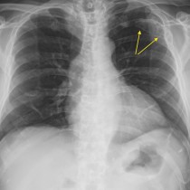
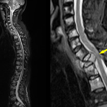
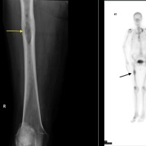
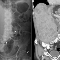
Myeloma
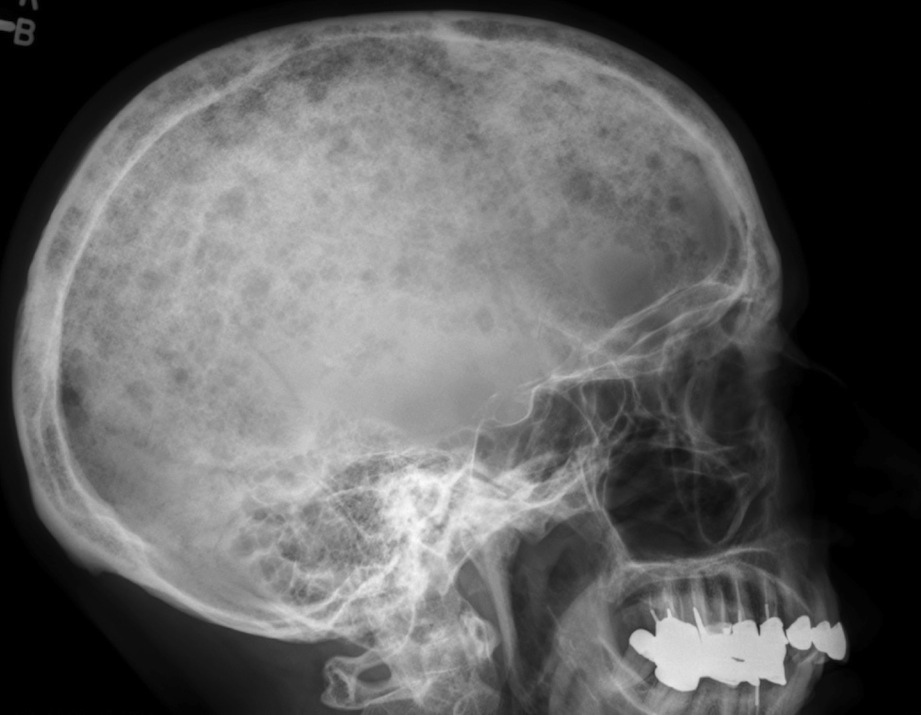
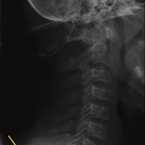
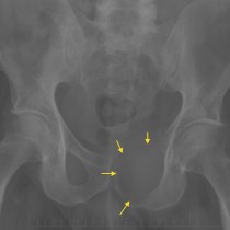
Myeloma. A lateral skull x-ray is routinely performed as part of a ‘skeletal survey’, when myeloma is suspected. This is the typical radiographic appearance of myeloma – multiple small lytic lesions. The other components of a skeletal survey are: cervical, thoracic and lumbar spine, both humeri, both femora, and the pelvis.