Myeloma in rib
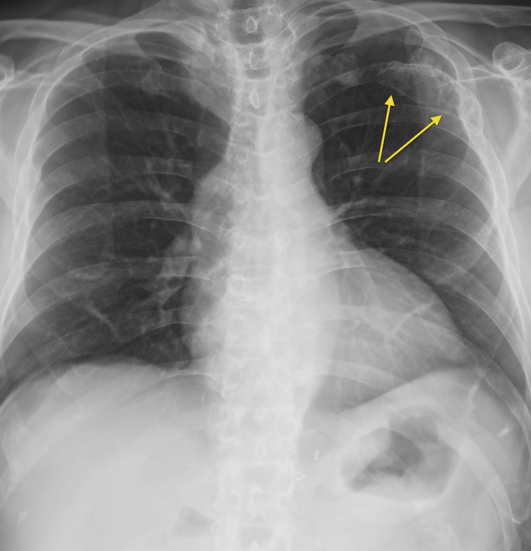
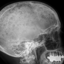
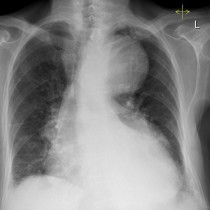
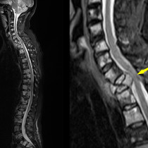
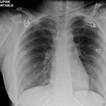
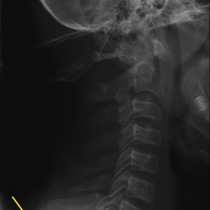
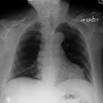
Here is a nice example of a lytic, expansile rib lesion in a patient with multiple myeloma (arrow). The chest radiograph is a component of the ‘skeletal survey’ typically carried out when someone is diagnosed with myeloma; the other radiographs performed as part of this are: lateral skull, cervical/thoracic/lumbar spine, humeri, femora and pelvis. In many centres, the traditional radiographic skeletal survey has been superseded by more sensitive imaging techniques such as whole body MRI.