• Close
    • Home
    • Videos
    • Tutorials
    • Gallery
    • Links
    • FAQs
    • All Case Studies
    • Diagnostic Imaging
      • Imaging Modalities
        • Radiography
        • Fluoroscopy
        • Ultrasound
        • CT
        • MRI
        • Nuclear Medicine
      • Contrast Media
      • Radiological Anatomy
      • Case Studies
      • PACS
      • Radiation Safety
      • Radiological Descriptive Terms
    • Interventional Radiology
      • Introduction to IR
      • Image-guided Biopsy
      • Case Studies
    • UCD Students
      • Weekly Rotation
      • Radiology Conferences
      • Resources
      • Undergraduate Medals
    • Electives
    • Careers
    • About Us
St. Vincent's University Hospital Radiology Department
  • Diagnostic Imaging
    • Imaging Modalities
      • Radiography
      • Fluoroscopy
      • Ultrasound
      • CT
      • MRI
      • Nuclear Medicine
    • Contrast Media
    • Radiological Anatomy
    • Case Studies
    • PACS
    • Radiation Safety
    • Radiological Descriptive Terms
  • Interventional Radiology
    • Introduction to IR
    • Image-guided Biopsy
    • Case Studies
  • UCD Students
    • Weekly Rotation
    • Radiology Conferences
    • Resources
    • Undergraduate Medals
  • Electives
  • Careers
  • About Us
  • Home
  • Videos
  • Tutorials
  • Gallery
  • Links
  • FAQs
  • All Case Studies

Malpositioned endotracheal tube

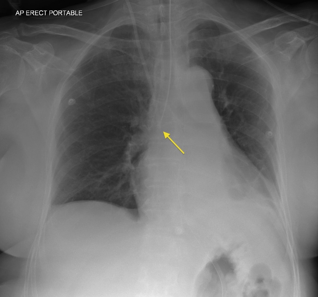
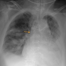
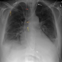
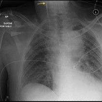
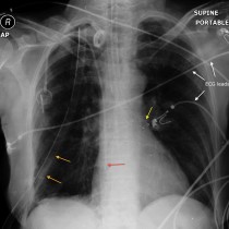
Malpositioned endotracheal tube

The tip of this ventilated patient’s endotracheal tubes several centimetres beyond the carina, in the right main bronchus (arrow). Note also the right internal jugular line.

Related Case Studies

  • Malpositioned ET tube
  • Spaghetti junction
  • Malpositioned subclavian line
  • Endotracheal tube at carina
  • Selective intubation of left main bronchus
  • Air bronchograms
Back
  • Interventional Radiology
  • Diagnostic Imaging
    • Emergencies
    • Neuro
    • Tubes & Lines
      • Malpositioned endotracheal tube
      • Malpositioned subclavian line
      • Biliary drainage catheter
      • Malpositioned endotracheal tube
      • Brachytherapy rods
      • Ventriculoperitoneal shunt
      • Ureteric stent
      • Atrial appendage closure device
      • Defibrillation pads – CXR
      • Malpositioned ET tube
      • NG tube coiled in oesophagus
      • EVAR
      • Ring pessary
      • Tubal ligation clips
      • Sengstaken Blakemore tube
      • Ureteric stent
      • Fractured central venous catheter
      • Pneumothorax from central line insertion
      • Intra-aortic balloon pump
      • Selective intubation of left main bronchus
      • Endotracheal tube at carina
      • Spaghetti junction
    • Trauma
    • Oncology
    • Hepatobiliary
    • Genitourinary
    • Gastrointestinal
    • Chest
    • Musculoskeletal
    • Vascular

All Case Studies

  • Main Categories

    • Interventional Radiology
      • Vascular IR
      • Non-Vascular IR
      • IR Oncology
      • Musculoskeletal IR
    • Diagnostic Imaging
      • Emergencies
      • Neuro
      • Tubes & Lines
      • Trauma
      • Oncology
      • Hepatobiliary
      • Genitourinary
      • Gastrointestinal
      • Chest
      • Musculoskeletal
      • Vascular
  • CONTACT US

    Department of Radiology
    St Vincent’s University Hospital
    Elm Park
    Dublin 4
    Send email

    Links

    • FAQs
    • Links

    SOCIAL

    © Copyright St Vincent's University Hospital Radiology Department 2025
    Web Design Dublin

    Search St. Vincent's University Hospital Radiology Department

    This website uses cookies. Accept Read More