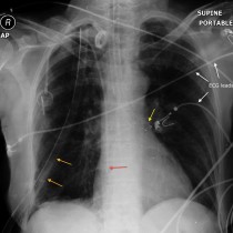
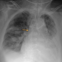
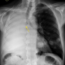
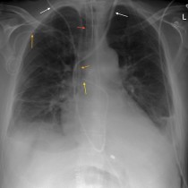
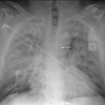
Endotracheal tube at carina
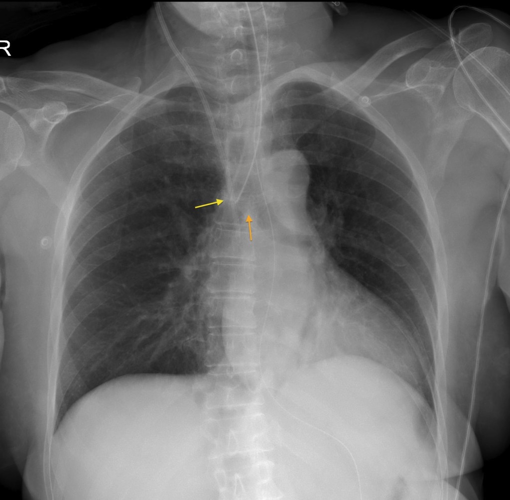
Endotracheal tube at carina. In this example, the tip of the ETT (yellow arrow) is too close to the carina (orange arrow), and is at the origin of the right main bronchus. In this position, there is a risk of overventilation of the right lung and underventilation of the left.