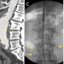
Calcified abdominal aortic aneurysm – lumbar spine radiograph
Not infrequently, we pick up incidental asymptomatic abdominal aortic aneurysms on lumbar spine radiographs. This is possible only if the wall of the aneurysm is calcified, which many of them are.
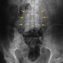
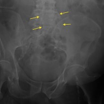
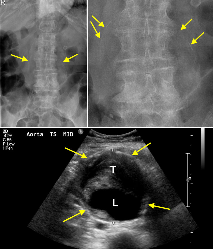
In the example shown here, from a 90 year old woman complaining of low back pain, curvilinear calcification is visible on either side of the lumbar spine (arrows in top left image – more clearly seen on the magnified image on the right). This calcification is in the wall of the aorta, which must therefore be aneurysmal to produce this bulging appearance.
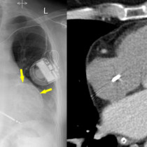
An ultrasound was recommended and confirmed that there was an aneurysm – the aorta measured up to 7.5 cm in diameter, placing the patient at high risk of rupture and death. The ultrasound image, below, shows the echogenic calcified wall of the aorta (arrows). The lumen of the aorta is irregular in shape and is the black (‘anechoic’) area indicated by the ‘L’. The remainder of the material inside the aortic wall represents chronic thrombus ‘T’, which is frequently present in aortic aneurysms (and can embolize to the lower limbs).
Other potential causes of incidental calcification on a lumbar spine radiograph include:
- gallstones
- renal stones
- calcified mesenteric lymph nodes (quite common, especially in elderly patients)
- calcified uterine fibroids in the pelvis
- appendicoliths
- iliac artery aneurysms
While in this case there was significant degenerative change in the lower lumbar spine which would explain the patient’s symptoms, it should be remembered that abdominal aortic aneurysms can present with chronic low back pain therefore identifying them on lumbar spine radiographs may not always be a truly ‘incidental’ finding.
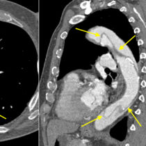
You can read about endovascular aneurysm repair (EVAR) here.