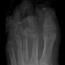
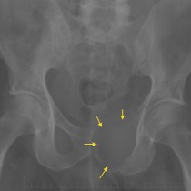
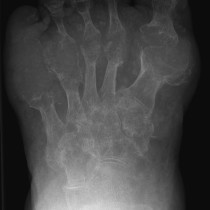
Arterial calcification in a diabetic
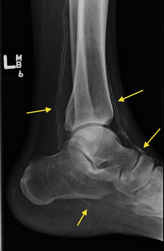
Arterial calcification in a diabetic. This patient is only 50 years old, but his anterior and posterior tibial arteries are heavily calcified (arrows). In a patient of this age, this finding should lead you to assume that he is diabetic (which he was). Other potential findings in the foot of a diabetic would include a neuropathic joint, and bone destruction due to osteomyelitis.