Uterine fibroid embolization
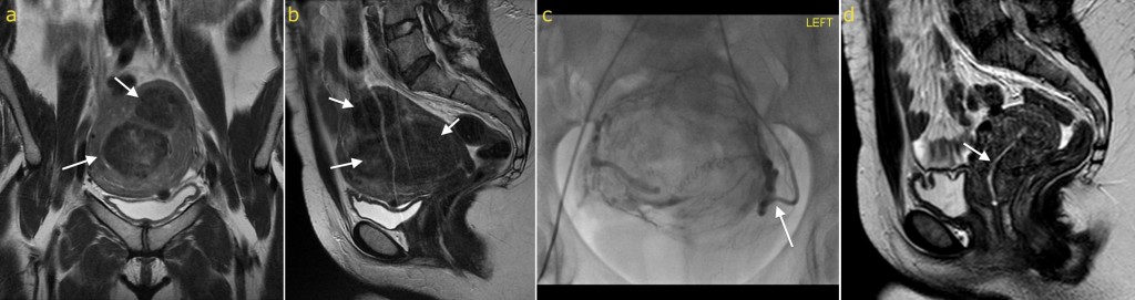
Uterine fibroid embolization (UFE). This 38 year old woman had severe menorrhagia caused by large uterine fibroids. On initial MRI the fibroids are visible as low signal masses in the myometrium on coronal (a) and sagittal (b) T2-weighted images (note the bright urine in the bladder). Uterine fibroid embolization is performed by advancing a catheter through one the femoral arteries and then manipulating it into the uterine arteries (which are branches of the internal iliac arteries). On image (c), contrast has been injected through the catheter in the left uterine artery (arrow) and delineates the blood supply to the fibroids. The arteries are occluded by injecting tiny (sub-millimetre) microspheres through the catheter. Follow-up MRI (d) performed three months post UFE shows that the fibroids have completely disappeared; note that the endometrium, which was distorted and obscured by the fibroids on the initial MRI, is now clearly visible (arrow). The patient was now asymptomatic.