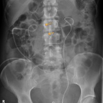
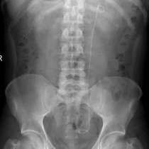
Tubal ligation clips
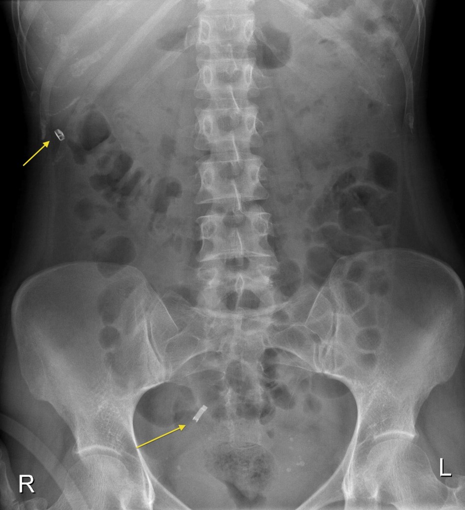
Tubal ligation clips. These large metallic staples are commonly seen in abdominal radiographs and are typical of tubal ligation clips. They commonly fall off the fallopian tubes and move around in the peritoneal cavity, as in this case where one of the clips is in the pelvis but the other is in the right upper quadrant.