Thyroid carcinoma
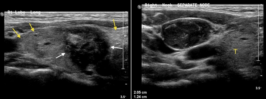
Thyroid carcinoma. This woman in her twenties was complaining of a firm lump in the right side of her neck. Ultrasound was requested and shows an irregularly-shaped hypoechoic nodule in the right lobe of her thyroid gland, suspicious for malignancy. The image on the left shows the normal hyperechoic thyroid tissue (yellow arrows), encompassing the hypoechoic lesion (white arrows). In addition, the image on the right shows an abnormal-looking lymph node adjacent to the thyroid. Fine needle aspiration (FNA) of the thyroid nodule was performed, and confirmed papillary carcinoma.