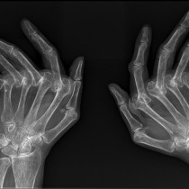
Psoriatic arthritis – DIP erosions
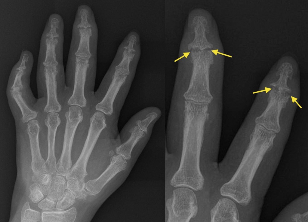
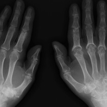
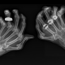
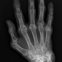
This is an example of the classic radiographic appearance of psoriatic arthritis, with marginal erosions of the distal interphalangeal joints (best appreciated on the magnified view of the second and third fingers, on the right, arrows). By ‘marginal’ erosions we mean erosions at the edge of the joint – the bone gets eroded here first because the overlying cartilage is much thinner than in the centre of the joint. You will notice how the PIP and MCP joints are spared, whereas in the classical appearance of rheumatoid arthritis (RA) we see involvement of the MCP and PIP joints with sparing of the DIP joints. In reality, this textbook pattern of psoriatic arthritis is not often seen, as there are several other potential patterns. For example, it often manifests as a symmetrical, proximal arthropathy involving the MCP and PIP joints, indistinguishable (radiographically) from RA. Another common feature of psoriatic arthritis is the sausage digit – note the diffuse swelling of the second, third and fourth fingers in this example.