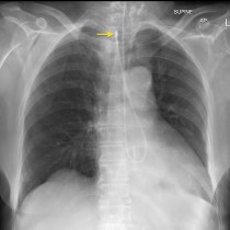
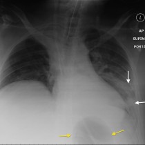
NG tube coiled in oesophagus
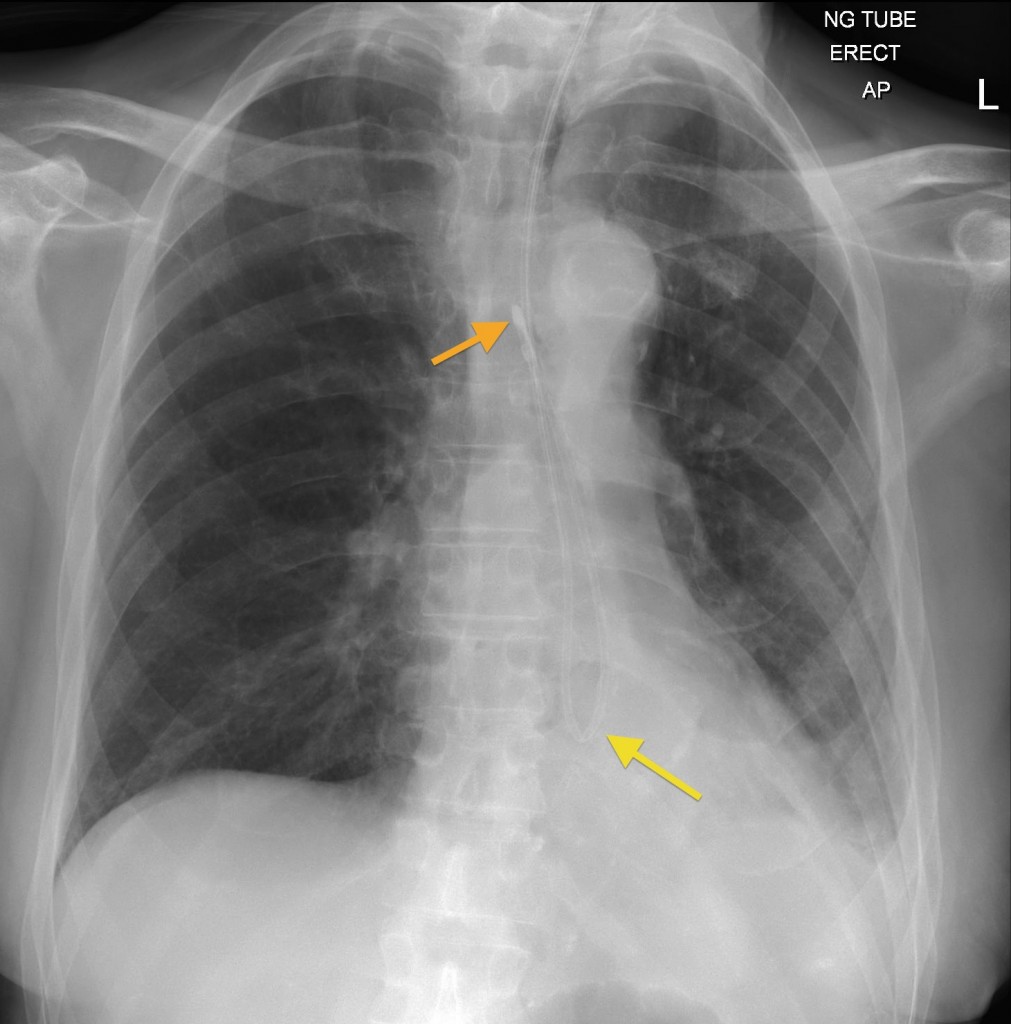
Malpositioned NG tube. This CXR was obtained to check the position of an NG tube that had just been inserted. The tube coils on itself in the distal oesophagus (yellow arrow) and the radio-opaque tip is actually located in the upper oesophagus (orange arrow), requiring repositioning.